A dental radiograph, more commonly known as an x-ray, is formed by a controlled burst of X-ray radiation which penetrates oral structures at different levels. Dental caries, infections and other changes in the bone density are visible on these x-rays. They are used to give dentists a better understanding of what is happening in areas that aren’t visible by examining the mouth.
Why do dentists take x-rays?
There are many different types of dental radiographs, from periapical, bitewings to panoramic. Each different type is designed to look at specific sections of the tooth/mouth.
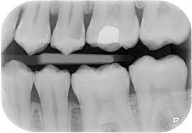 Bitewing X-rays show the upper and lower back teeth and how the teeth touch each other in a single view. These X-rays are used to check for decay between the teeth and to show how well the upper and lower teeth line up. They also show a bone loss when severe gum disease or a dental infection is present.
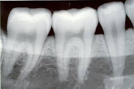 Periapical X-rays show the entire tooth, from the exposed crown to the end of the root and the bones that support the tooth. These X-rays are used to find dental problems below the gum line or in the jaw, such as impacted teeth, abscesses, cysts, tumours, and bone changes linked to some diseases.
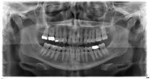 Panoramic X-rays show a broad view of the jaws, teeth, sinuses, nasal area, and temporomandibular (jaw) joints. These X-rays do not find cavities. These X-rays do show problems such as impacted teeth, bone abnormalities, cysts, solid growths (tumours), infections, and fractures.
How often do we need to take x-rays?
Bitewing x-rays are generally taken on a patient’s first visit for a check-up and clean and every two years after that to monitor any tooth decay. Other x-rays are taken on patients when they are required depending on different circumstances.